Sugar- and Artificially Sweetened Beverages and the Risks of Incident Stroke and Dementia

Stroke. 2017 Apr 20. pii: STROKEAHA.116.016027. doi: 10.1161/STROKEAHA.116.016027. [Epub ahead of print]

Sugar- and Artificially Sweetened Beverages and the Risks of Incident Stroke and Dementia: A Prospective Cohort Study.

Pase MP1, Himali JJ2, Beiser AS2, Aparicio HJ2, Satizabal CL2, Vasan RS2, Seshadri S2, Jacques PF2.

Abstract

BACKGROUND AND PURPOSE:

Sugar- and artificially-sweetened beverage intake have been linked to cardiometabolic risk factors, which increase the risk of cerebrovascular disease and dementia. We examined whether sugar- or artificially sweetened beverage consumption was associated with the prospective risks of incident stroke or dementia in the community-based Framingham Heart Study Offspring cohort.

METHODS:

We studied 2888 participants aged >45 years for incident stroke (mean age 62 [SD, 9] years; 45% men) and 1484 participants aged >60 years for incident dementia (mean age 69 [SD, 6] years; 46% men). Beverage intake was quantified using a food-frequency questionnaire at cohort examinations 5 (1991-1995), 6 (1995-1998), and 7 (1998-2001). We quantified recent consumption at examination 7 and cumulative consumption by averaging across examinations. Surveillance for incident events commenced at examination 7 and continued for 10 years. We observed 97 cases of incident stroke (82 ischemic) and 81 cases of incident dementia (63 consistent with Alzheimer’s disease).

RESULTS:

After adjustments for age, sex, education (for analysis of dementia), caloric intake, diet quality, physical activity, and smoking, higher recent and higher cumulative intake of artificially sweetened soft drinks were associated with an increased risk of ischemic stroke, all-cause dementia, and Alzheimer’s disease dementia. When comparing daily cumulative intake to 0 per week (reference), the hazard ratios were 2.96 (95% confidence interval, 1.26-6.97) for ischemic stroke and 2.89 (95% confidence interval, 1.18-7.07) for Alzheimer’s disease. Sugar-sweetened beverages were not associated with stroke or dementia.

CONCLUSIONS:

Artificially sweetened soft drink consumption was associated with a higher risk of stroke and dementia.

Citation

http://stroke.ahajournals.org/content/early/2017/04/20/STROKEAHA.116.016027

© 2017 American Heart Association, Inc.
 

Risk of Pneumonia Associated with Incident Benzodiazepine Use Among Adults with Alzheimer Disease

CMAJ. 2017 Apr 10;189(14):E519-E529. doi: 10.1503/cmaj.160126.

Risk of pneumonia associated with incident benzodiazepine use among community-dwelling adults with Alzheimer disease.

Taipale H1, Tolppanen AM2, Koponen M2, Tanskanen A2, Lavikainen P2, Sund R2, Tiihonen J2, Hartikainen S2.

Abstract

Background

Knowledge regarding whether benzodiazepines and similarly acting non-benzodiazepines (Z-drugs) are associated with an increased risk of pneumonia among older adults is lacking. We sought to investigate this association among community-dwelling adults with Alzheimer disease, a condition in which both sedative/hypnotic use and pneumonia are common.

Methods

We obtained data on all community-dwelling adults with a recent diagnosis of Alzheimer disease in Finland (2005-2011) from the Medication use and Alzheimer disease (MEDALZ) cohort, which incorporates national registry data on prescriptions, reimbursement, hospital discharges and causes of death. Incident users of benzodiazepines and Z-drugs were identified using a 1-year washout period and matched with nonusers using propensity scores. The association with hospital admission or death due to pneumonia was analyzed with the Cox proportional hazards model and adjusted for use of other psychotropic drugs in a time-dependent manner.

Results

Among 49 484 eligible participants with Alzheimer disease, 5232 taking benzodiazepines and 3269 taking Z-drugs were matched 1:1 with those not taking these drugs. Collectively, use of benzodiazepines and Z-drugs was associated with an increased risk of pneumonia (adjusted hazard ratio [HR] 1.22, 95% confidence interval [CI] 1.05-1.42). When analyzed separately, benzodiazepine use was significantly associated with an increased risk of pneumonia (adjusted HR 1.28, 95% CI 1.07-1.54), whereas Z-drug use was not (adjusted HR 1.10, 95% CI 0.84-1.44). The risk of pneumonia was greatest within the first 30 days of benzodiazepine use (HR 2.09, 95% CI 1.26-3.48).

Interpretation

Benzodiazepine use was associated with an increased risk of pneumonia among patients with Alzheimer disease. Risk of pneumonia should be considered when weighing the benefits and risks of benzodiazepines in this population.

Citation

http://www.cmaj.ca/content/189/14/E519

© 2017 Canadian Medical Association or its licensors.


Association between Midlife Vascular Risk Factors and Estimated Brain Amyloid Deposition

JAMA. 2017 Apr 11;317(14):1443-1450. doi: 10.1001/jama.2017.3090.

Association Between Midlife Vascular Risk Factors and Estimated Brain Amyloid Deposition.

Gottesman RF1, Schneider AL2, Zhou Y3, Coresh J4, Green E5, Gupta N6, Knopman DS7, Mintz A8, Rahmim A3, Sharrett AR4, Wagenknecht LE9, Wong DF10, Mosley TH11.

Abstract

Importance

Midlife vascular risk factors have been associated with late-life dementia. Whether these risk factors directly contribute to brain amyloid deposition is less well understood.

Objective

To determine if midlife vascular risk factors are associated with late-life brain amyloid deposition, measured using florbetapir positron emission tomography (PET).

Design, Setting, and Participants

The Atherosclerosis Risk in Communities (ARIC)-PET Amyloid Imaging Study, a prospective cohort study among 346 participants without dementia in 3 US communities (Washington County, Maryland; Forsyth County, North Carolina; and Jackson, Mississippi) who have been evaluated for vascular risk factors and markers since 1987-1989 with florbetapir PET scans in 2011-2013. Positron emission tomography image analysis was completed in 2015.

Exposures

Vascular risk factors at ARIC baseline (age 45-64 years; risk factors included body mass index ≥30, current smoking, hypertension, diabetes, and total cholesterol ≥200 mg/dL) were evaluated in multivariable models including age, sex, race, APOE genotype, and educational level.

Main Outcomes and Measures

Standardized uptake value ratios (SUVRs) were calculated from PET scans and a mean global cortical SUVR was calculated. Elevated florbetapir (defined as a SUVR >1.2) was the dependent variable.

Results

Among 322 participants without dementia and with nonmissing midlife vascular risk factors at baseline (mean age, 52 years; 58% female; 43% black), the SUVR (elevated in 164 [50.9%] participants) was measured more than 20 years later (median follow-up, 23.5 years; interquartile range, 23.0-24.3 years) when participants were between 67 and 88 (mean, 76) years old.

Elevated body mass index in midlife was associated with elevated SUVR (odds ratio [OR], 2.06; 95% CI, 1.16-3.65).

At baseline, 65 participants had no vascular risk factors, 123 had 1, and 134 had 2 or more; a higher number of midlife risk factors was associated with elevated amyloid SUVR at follow-up (30.8% [n = 20], 50.4% [n = 62], and 61.2% [n = 82], respectively).

In adjusted models, compared with 0 midlife vascular risk factors, the OR for elevated SUVR associated with 1 vascular risk factor was 1.88 (95% CI, 0.95-3.72) and for 2 or more vascular risk factors was 2.88 (95% CI, 1.46-5.69).

No significant race × risk factor interactions were found. Late-life vascular risk factors were not associated with late-life brain amyloid deposition (for ≥2 late-life vascular risk factors vs 0: OR, 1.66; 95% CI, 0.75-3.69).

Conclusions and Relevance

An increasing number of midlife vascular risk factors was significantly associated with elevated amyloid SUVR; this association was not significant for late-life risk factors. These findings are consistent with a role of vascular disease in the development of Alzheimer disease.

Citation

http://jamanetwork.com/journals/jama/article-abstract/2616396

© 2017 American Medical Association. All Rights Reserved.


The Discovery of Alzheimer’s Disease

Dialogues Clin Neurosci. 2003 Mar; 5(1): 101–108.

The discovery of Alzheimer’s disease

Hanns Hippius, MD*
Hanns Hippius, Psychiatrische Klinik der LMU, Munich, Germany;

Gabriele Neundörfer, MD
Gabriele Neundörfer, Psychiatrische Klinik der LMU, Munich, Germany;

Abstract

On Novembers, 1306, a clinical psychiatrist and neuroanatomist, Alois Alzheimer, reported “A peculiar severe disease process of the cerebral cortex” to the 37th Meeting of South-West German Psychiatrists in Tubingen, He described a 50-year-old woman whom he had followed from her admission for paranoia, progressive sleep and memory disturbance, aggression, and confusion, until her death 5 years later. His report noted distinctive plaques and neurofibrillary tangles in the brain histology. It excited little interest despite an enthusiastic response from Kraepelin, who promptly included “Alzheimer’s disease” in the 3ih edition of his text Psychiatrie in 1910. Alzheimer published three further cases in 1909 and a “plaque-only” variant in 1911, which reexamination of the original specimens in 1993 showed to be a different stage of the same process, Alzheimer died in 1915, aged 51, soon after gaining the chair of psychiatry in Breslau, and long before his name became a household word.

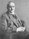
The 37th Meeting of South-West German Psychiatrists (37 Versammlung Sudwesideutscher Irrenarzte) was held in Tubingen on November 3, 1906. At the meeting, Alois Alzheimer (Figure 1), who was a lecturer(Privatdozent) at the Munich University Hospital and a coworker of Emil Kraepelin, reported on an unusual case study involving a “peculiar severe disease process of the cerebral cortex” (Uber einen eigenartigen, schweren Erkrankungsprozeβ der Hirnrinde).


Figure 1.

Figure 1. Alois Alzheimer. About 1909. © Archive for History of Psychiatry, Department of Psychiatry University of Munich. With permission.

Prelude

Alzheimer described the long-term study of the female patient Auguste D., whom he had observed and investigated at the Frankfurt Psychiatric Hospital in November 1901 , when he was a senior assistant, there. Alzheimer had been interested in the symptomatology, progression, and course of the illness of Auguste D. from the time of her admission, and he documented the development of her unusual disease very precisely from the beginning.

In March 1901 , the husband of the 50-year-old woman had noticed an untreatable paranoid symptomatology in his wife and then - in fast progression and with increasing intensity - sleep disorders, disturbances of memory, aggressiveness, crying, and progressive confusion. Eventually, the husband was forced to take his wife to the Community Psychiatric Hospital at Frankfurt am Main, lite symptomatology increasingly deteriorated and so Auguste D. remained an inpatient of the hospital up to her death on April 8, 1906.

After the autopsy, Alzheimer was able to investigate the brain of Auguste D.both morphologically and histologically. These results and their relationship with the clinical findings recorded over more than 4 years were the basis for Alzheimer’s lecture at the Tubingen meeting.

The chairman of the session was the very prominent psychiatrist from the University of Freiburg, Alfred Hoche (1865-1943). Hoche was a scientific opponent of Kraepelin and his nosological concept and classification of psychiatric diseases. Kraepelin was not in the audience during Alzheimer’s presentation.

After Alzheimer’s lecture, Hoche, departing from the usual role of a chairman, did not comment on Alzheimer’s presentation and only once or twice asked the audience for comments or questions. He stated that, there was no need for discussion and invited the next, speakers to continue with their lectures. These were two contributions to psychoanalytical topics, and were followed by long and very lively discussions, including some active comments from the chairman.

The lack of interest from the numerous and well-known scientists in the audience was a great disappointment, for Alzheimer. Moreover, only a very short abstract, was printed in the official proceedings of the meeting.1 Tubingen’s public press commented extensively on the psychoanalytical lectures, whereas only two lines were devoted to Alzheimer’s lecture. Such was the beginning of communication on research into Alzheimer’s disease!2

Alois Alzheimer

Alois Alzheimer was born into a Catholic family on June 14, 1864, in the small town of Marktbreit in Lower Frankonia close to Würzburg on the river Main.2-4 His father was a royal notary in the Kingdom of Bavaria who had lost his first, wife 2 years previously to puerperal fever after giving birth to their first son. Alzheimer’s father married the sister of his dead wife and had six more children with her - the eldest child was Alois Alzheimer.

Alois Alzheimer went to elementary school in Marktbreit and later to classic secondary school in Aschaffenburg. After he left, school, Alzheimer became a college student in Berlin, Freiburg, and Wtirzburg (1883-1885). Very early on, in the first few academic trimesters, he became interested in anatomy and learned to work with microscopes.

As a young student, he attended some lectures on forensic psychiatry, but later during clinical training he was extensively occupied in all clinical disciplines, with one notable exception: he probably never attended clinical lectures in psychiatry! After a dissertation in anatomy, he finished his studies at Wtirzburg and obtained the official diploma in medicine with magna cum laude.

At this time, there were no indications that Alzheimer was destined to follow a career in psychiatry. However, a more or less accidental event after the end of his studies in medicine may have influenced him in this direction. At the end of the 19th century, some very wealthy German families had an unusual approach to the care of a mentally ill relative: they engaged a young medical doctor to travel with the patient. Alzheimer had such an offer and traveled for 5 months (May to October 1888) with a mentally ill female patient. Unfortunately, no information is known regarding this patient’s illness or identity, or the travel itinerary.

Upon returning from this journey, at the age of 24 years, Alzheimer applied for a position as assistant in the Community Hospital for Mental and Epileptic Patients (Städlische Anstalt fur Irre und Epileptiker) in Frankfurt am Main. The director of the Frankfurt Hospital, Emil Sioli (1852-1922), accepted Alzheimer’s application. Alzheimer worked with Sioli for 15 years (1888-1903) and was strongly influenced by him: Alzheimer thus became an all-round skilled clinician.

Sioli had held this position in Frankfurt since 1888 and he was the successor of the pediatrician H. Hoffmann (well known as the author of Shock-Headed Peter [Der StruwwelpeterJ).As a psychiatrist, Sioli directed the hospital with the main idea of nonrestraint psychiatry, an idea introduced from Great Britain, but at that time still controversial in Germany.

Today, many people believe that Alzheimer was a pure neuropathologist, but all information on his own selfassessment, as well as the summary of his scientific publications - after working with Sioli - demonstrate that he identified himself primarily as a clinical psychiatrist responsible for patients.

In addition to his development, as a clinician in Frankfurt, Alzheimer did not neglect. Ms interest in anatomy dating from his time as a young student in Berlin and Würzburg. This interest was enhanced by Franz Nissl (1860-1919), who came from Munich to work with Sioli in. Frankfurt 1 year after Alzheimer.

Nissl had already worked in neuroanatomy and neuropathology as a student, and had discovered a special histological staining technique (Nissl stain), which is still in use today. In Munich, Nissl had been a coworker of B. von Guddcn in his brain research laboratory. After the tragic death of von Gudden, who was found drowned with his patient Ludwig II, King of Bavaria, in 1886, Nissl sought, a new comparable position and, with the help of Sioli, became senior assistant at the Frankfurt Hospital in 1889.

From the beginning, Nissl and Alzheimer became good colleagues and close friends. The more senior Nissl encouraged young Alzheimer to actively continue research alongside his clinical work. Alzheimer followed Nissl’s advice and worked on topics such as general paresis in children and young adults,5 and brain atrophy in patients with cerebral arteriosclerosis,6 epilepsy,7 or dementia] diseases.8 He also published pioneering ideas on the contribution of the cortex to pathology, as the anatomical basis of some psychotic diseases.9

During his Frankfurt years, in 1895, Alzheimer married the very wealthy Cecilia Geisenheimer (née Wallerstein); Nissl was a witness at the marriage ceremony. Due to the prosperous financial background of his wife, Alzheimer was henceforth financially independent. His aim was to become an independent clinical director of a psychiatric hospital in which he could do research, but not exclusively Nissl left Frankfurt in 1896 because he had been invited by Emil Kraepelin (1856-1926) to work at the University Hospital of Heidelberg, which was directed by Kraepelin between 1890 and 1903.

Nissl accepted the invitation because he thus achieved a position at a university with better conditions for research. Both Nissl and Alzheimer regretted that they could no longer work together at the same hospital. However, they continued their friendship and their scientific exchange for the rest of their lives.

On the other hand, Nissl’s move to Heidelberg brought about an improvement in Alzheimer’s position at the Frankfurt Hospital. Sioli recommended Alzheimer to the authorities as Nissl’s successor as first assistant and deputy director of the hospital.

The official appointment to this position was in July 1896. This appointment represented an important step for Alzheimer toward his professional target: to become the director of a psychiatric hospital.

The following years were satisfactory for Alzheimer not only with regard to his professional situation, but also with respect, to his particularly harmonious family life with his wife and three children born between 1895 and 1900.2

1901

For Alzheimer, the year 1901 marked a difficult turning point in his life. Some months after the birth of their third child, his 41 -year-old wife died. Alzheimer was now a widower and had to take care of three children. Although his income from Ms position at the hospital was small, he had his wife’s extensive inheritance. One of his unmarried sisters moved to Frankfurt to look after the household because Alzheimer wanted to live with his family and to work at, or near to, Frankfurt. He planned to apply for leading positions in this area.

To overcome the grief of his wife’s death, Alzheimer worked more intensively at the hospital than ever before. He saw all newly admitted patients and made a detailed and extensive documentation of his findings. On November 26, 1901, he investigated the newly admitted female patient Auguste D., not imagining for one moment, that the clinical investigation of this patient would be the starting point for a development that would make him famous throughout the world!

From Frankfurt to Munich via Heidelberg

Apart from his very intensive clinical work, Alzheimer - together with Sioli - organized the establishment of a special branch hospital for mental patients close to Frankfurt in the Taunus mountains. In addition, he began to write a so-called Habilitationssdirift (postdoctoral thesis for a university lecturing qualification) as a basis for an application at a medical faculty of a German university.

He was in possession of the clinical and the postmortem findings of 320 patients with the diagnosis of “Progressive Paralyse” (general paresis), investigated at the Frankfurt Hospital since 1888. (Around 1900, more than 25% of chronic psychiatric inpatients suffered from this disease and were hospitalized up to their death. The relationship between syphilis and general paresis was still controversial: Treponema pallidum, [Spirochaela pallida] had not yet been discovered and no effective treatment was available.)

In the summer of 1902, little more than one year after the death of Alzheimer’s wife, Emil Kraepelin invited him to join the Heidelberg research team as assistant to the Heidelberg Hospital. This was a great honor because Kraepelin was at the time one of the most, prominent and influential psychiatrists in Germany.

In addition, Alzheimer’s great friend Nissl had then been working in the Heidelberg Hospital for 7 years. In spite of many reasons in favor of Heidelberg, Alzheimer refused Kraepelin’s invitation and applied - unsuccessfully - for the leading position in a Hessian state hospital.

When Nissl heard about, this, he persuaded Kraepelin to repeat his offer of a position at the Heidelberg Hospital to Alzheimer. Kraepelin did so and Alzheimer accepted; he moved to Heidelberg at the end of 1902.10

Sioli and the Frankfurt, authorities explicitly regretted the departure of Alzheimer. However, Sioli approved of Alzheimer’s decision, since it led to a university position (the University of Frankfurt was only established in 1914). Sioli promised Alzheimer that he would tell him of the fate of all the patients who had been of special interest to Alzheimer from a scientific point of view. Thus, some years later, Alzheimer obtained information on the course of Auguste D.’s illness and her death at the Frankfurt Hospital in April 1906.

Alzheimer moved to Heidelberg expecting to work there for a long time. However, just one month later in April 1903, the Professor of Psychiatry in Munich, A. Bumm (1849-1903), died at the age of 54. For some years, Bumm had been responsible for the planning and construction of a new modern, large university hospital for psychiatry. At the time of Bumm’s unexpected death, the building was not yet finished and the Munich chair suddenly became vacant. On the recommendation of the Faculty of Medicine, the chair and directorship were offered to Kraepelin.

After only momentary hesitation, Kraepelin agreed to soon take up the position and moved in autumn 1902. He was accompanied by three coworkers from his Heidelberg team, one of whom was Alzheimer. Kraepelin used the remaining year till the official opening of the hospital in November 1904 to work on his textbooks and undertook a long voyage to explore Indonesia.

During this time, Alzheimer’s task in Munich was the supervision of the completion of the building and the organization of hospital equipment. After his return, Kraepelin stated that. Alzheimer had done an excellent job.10 With regard to hospital equipment, a very modern and spacious histopathological laboratory with the most modem microscopes and other apparatus was established (Figure 2), enabling Alzheimer to continue his histopathological research.

Figure 2.

Figure 2. Alzheimer’s modern histopathological laboratory in the Psychiatric University Hospital in Munich, 1904. © Archive for History of Psychiatry, Department of Psychiatry University of Munich. With permission.

After the opening of the hospital in November 1904, R. Gaupp (1870-1953) (Figure 3) was appointed senior assistant and Alzheimer became Kraepelin’s first, research assistant. In this position, Alzheimer received no payment, but he could devote all his time to research. Alzheimer’s remarkable private fortune enabled him to work under these peculiar conditions.

Figure 3.

Figure 3. (Left to right) A. Alzheimer, E. Kraepelin, R. Gaupp, and F. Nissl. About 1906. © Archive for History of Psychiatry, Department of Psychiatry University of Munich. With permission.

Alzheimer was head of the histopathological laboratory until 1912. During these 8 years, numerous young scientists from many countries were trained by Alzheimer and later became famous neuropathologists or clinical psychiatrists. The list, of Alzheimer’s coworkers (Figure 4) includes many prominent names - N. Achucarro, I. Bonfiglio, L. Casamaior, U. Cerletti, H-G Crcutzfeld, C. v. Econome, A. Jakob, K. Kleist, F. H. Lewy, L. Merzbacher, G. Perusini, and W. Spielmeyer - a who’s who of contemporary neuropathology!

Figure 4.

Figure 4. Alzheimer and coworkers in Munich. Back (left to right); F. Lotmar; unknown; St Rosental; Allers (?); unknown; A. Alzheimer; M. Achucarro, F H. Levy. Front (left to right); Frau Grombach; U. Cerletti; unknown; F Bonfiglio; G. Perusini. About 1909.

In October 1903, a short time after moving to Munich, Alzheimer arranged for his children to follow him and they all lived in a large house near the hospital, together with his sister as housekeeper. Furthermore, at the end of 1904, he bought, a big weekend house beside a small lake near Munich.

An important step in Alzheimer’s academic career came in November 1903 when he presented hisHabilitationsschrift in Munich. ‘Ill e manuscript, entitled Differential diagnosis of general paresis on the basis of histological studies (Histologische Studien zur Differ entialdiagnose der progressiven Paralyse) , was printed as an almost 300-page book soon afterwards 11and Alzheimer was appointed Privatdozent (lecturer) in August 1904.

Discovery

The case of Auguste D.

After the Munich Hospital had opened (November 11, 1904), Alzheimer hoped to again have more time for his research. This happened only for a short time, but with great effect. In April 1906, Sioli, with whom Alzheimer worked in .Frankfurt, informed him of the death of the patient Auguste D., arranged an autopsy, and gave him brain material for investigation. By this means, epoch-making research was enabled.12,13

Alzheimer discovered and described the histological alterations later known as plaques and neurofibrillary tangles.14 He presented these findings to Kraepelin and the other scientists in the Munich research team, convincing all of them that, such histopathological findings in connection with such a clinical symptomatology and course of illness had never been seen before.

Kraepelin encouraged Alzheimer to present the case of Auguste D. as soon as possible at the next scientific congress of German psychiatrists in the autumn of 1906 in Tubingen. The lack of response to this discovery at this meeting was very disappointing for Alzheimer, but he did not give up his search for comparable cases. He felt, satisfied that his lecture, which had not been mentioned at Tubingen, was published one year after the conference.15

Due to changes at the Munich Hospital, Alzheimer’s hopes of being able to devote all his time to research in the histopathological laboratory were dashed. Robert Gaupp, who had moved together with Kraepelin and Alzheimer from Heidelberg to Munich, was offered the chair of psychiatry and the directorship of the Medical Faculty of the University of Tubingen (1906-1939). Gaupp accepted this appointment and left Munich in October 1906. Kraepelin entrusted Alzheimer, as Gaupp’s successor, with the position of deputy director.

Alzheimer was now occupied with many additional obligations: care of patients, training of young psychiatrists, teaching of students, expert reports in psychiatry, and administrative duties. Therefore, Alzheimer delegated the research in the histopathological laboratory to his team of coworkers, which every year was becoming bigger.

Notably, Gactano Perusini from Italy specialized in research on cases with dementing processes. After 1906, Perusini and Alzheimer observed three additional cases comparable to that of Auguste D., and Perusini published these four cases, together with all clinical and histopathological details in 1909.16

Between 1906 and 1909, Kraepelin prepared the 8th edition of his famous textbook Psychiatrie A As he had soon recognized the fundamental significance of Alzheimer’s findings, he included a report, on the case history of Auguste D. in the written text of 1908 and proposed calling this peculiar illness Alzheimer’s disease. Both volumes of the new edition of Kraepelin ‘s textbook came out in 1910.

In this way, very soon after the description of the first case, the diagnostic term Alzheimer’s disease was introduced by Kraepelin’s authority and, since that time, has been generally used. However, in spite of this fact, because this disease - presenile dementia with some unusual histological signs (plaques and neurofibrillary tangles) - was very rare, the name of Alois Alzheimer was almost forgotten for more than 50 years. During the last, few decades, the situation has changed considerably.

The case of Josef F.

In 1911, Alzheimer himself published again in a broader context on presenile and senile dementing processes.18 He described how the male patient Josef F. died after 3 years of hospitalization in Munich in 1910. Kraepelin had already mentioned the case of Josef F. in his textbook, and had diagnosed him as having Alzheimer’s disease17 before death. The histological investigation confirmed the clinical diagnosis, but there was one important difference. Alzheimer noticed that there were no neurofibrillary tangles in the slide preparations of Josef F.’s brain, only plaques.

For a long time, it was considered to be contradictory if “plaque-only” cases belonged to the same category as cases with plaques and neurofibrillary tangles. A singular situation in research in recent years has provided a solution to this problem. In 1995, after an intensive search of the Frankfurt archives, K. Maurer discovered the documentation of the clinical findings of Auguste D.19

Histopathological slide preparations of her brain were subsequently found in the Munich Institute of Neuropathology. Documentation on the illness of Josef F. up to his death was found in clinical archives of the Munich Psychiatric Hospital and, after a long search, M. B. Graeber finally discovered the brain slide preparation in the depot of the Munich Institute of Neuropathology, where it had been stored since 1911.19

The material of both cases (Auguste D. and Josef F.) was reinvestigated with modern ncurohistochcmical techniques. The results of this investigation and analysis of all findings together with a summary on literature and conceptual interpretations were published by H-J. Moller and M. B. Graeber.2

Their conclusion was that plaque-only cases and cases with plaques and neurofibrillary tangles are simply different stages in the development of the same disease process.20 This means that - in addition to his pioneering discovery of the case of Auguste D. in 1906 - a few years later, Alzheimer was the first person to describe an important stage of development of the illness associated with his name with the case of Josef F. also.

From Munich to Breslau

Kraepelin had promised Alzheimer that, after the departure of Gaupp (1906), he would only be in charge as deputy director for a short time. However, this state of affairs lasted 3 years until E. Rtidin (1874-1952) was appointed to take over all of Alzheimer’s routine duties. Since he now had more time for research, Alzheimer was mainly occupied from 1909 onwards with histopathological studies on all kinds of psychotic mental diseases, including dementia praccox (schizophrenia) and manicdepressive psychoses. The aim was to also find a neuropath ological basis for these so-called endogenous psychoses.

Kraepelin was especially hopeful that Alzheimer would be successful, in order to demonstrate that his concept of dichotomy of these psychotic diseases was right. Alzheimer intended to publish all his findings in a comprehensive book, but he was not able to finish this project. He was also occupied with more general problems of research in psychiatric illness, notably with the difficulties in correlating clinical diagnosis and postmortem findings.21

In addition, on Kraepclin’s advice, in 1910, together with 3 the neurologist M.Lewandowsky (1876-1918), Alzheimer established a new scientific journal Zeitschrift für die gesamte Neurologie und Psychiatrie. The first introductory contribution of this new journal was written by Alzheimer himself.21

In 1912, he was appointed Chair of Psychiatry at the University of Breslau. This position was the realization of his dreams as a young assistant at the psychiatric hospital at Frankfurt, for his professional life: to work as clinician and director responsible for a psychiatric hosr pital. Unfortunately, he had very few years left to work in Breslau, for he died there at the age of 51 on December 19,1915.

Citation
http://www.ncbi.nlm.nih.gov/pmc/articles/PMC3181715/

National Center for Biotechnology Information, U.S. National Library of Medicine


Physical Exercise as a Preventive or Disease-Modifying Treatment of Dementia and Brain Aging

Mayo Clin Proc. 2011 Sep;86(9):876-84. doi: 10.4065/mcp.2011.0252.

Physical exercise as a preventive or disease-modifying treatment of dementia and brain aging.

Ahlskog JE1, Geda YE, Graff-Radford NR, Petersen RC.

Abstract

A rapidly growing literature strongly suggests that exercise, specifically aerobic exercise, may attenuate cognitive impairment and reduce dementia risk. We used PubMed (keywords exercise and cognition) and manuscript bibliographies to examine the published evidence of a cognitive neuroprotective effect of exercise.

Meta-analyses of prospective studies documented a significantly reduced risk of dementia associated with midlife exercise; similarly, midlife exercise significantly reduced later risks of mild cognitive impairment in several studies.

Among patients with dementia or mild cognitive impairment, randomized controlled trials (RCTs) documented better cognitive scores after 6 to 12 months of exercise compared with sedentary controls.

Meta-analyses of RCTs of aerobic exercise in healthy adults were also associated with significantly improved cognitive scores.

One year of aerobic exercise in a large RCT of seniors was associated with significantly larger hippocampal volumes and better spatial memory; other RCTs in seniors documented attenuation of age-related gray matter volume loss with aerobic exercise.

Cross-sectional studies similarly reported significantly larger hippocampal or gray matter volumes among physically fit seniors compared with unfit seniors.

Brain cognitive networks studied with functional magnetic resonance imaging display improved connectivity after 6 to 12 months of exercise. Animal studies indicate thatexercise facilitates neuroplasticity via a variety of biomechanisms, with improved learning outcomes. Induction of brain neurotrophic factors byexercise has been confirmed in multiple animal studies, with indirect evidence for this process in humans.

Besides a brain neuroprotective effect, physical exercise may also attenuate cognitive decline via mitigation of cerebrovascular risk, including the contribution of small vessel disease to dementia.

Exercise should not be overlooked as an important therapeutic strategy.

Citation

http://www.ncbi.nlm.nih.gov/pubmed/21878600


Prolonged Sleep Duration as Marker of Early Neurodegeneration Predicting Dementia

Neurology. 2017 Feb 22. pii: 10.1212/WNL.0000000000003732. doi:[Epub ahead of print]

Prolonged sleep duration as a marker of early neurodegeneration predicting incident dementia.

Westwood AJ1, Beiser A1, Jain N1, Himali JJ1, DeCarli C1, Auerbach SH1, Pase MP2, Seshadri S2.

Abstract

OBJECTIVE

To evaluate the association between sleep duration and the risk of incident dementia and brain aging.

METHODS

Self-reported total hours of sleep were examined in the Framingham Heart Study (n = 2,457, mean age 72 ± 6 years, 57% women) as a 3-level variable: <6 hours (short), 6-9 hours (reference), and >9 hours (long), and was related to the risk of incident dementia over 10 years, and cross-sectionally to total cerebral brain volume (TCBV) and cognitive performance.

RESULTS

We observed 234 cases of all-cause dementia over 10 years of follow-up. In multivariable analyses, prolonged sleep duration was associated with an increased risk of incident dementia (hazard ratio [HR] 2.01; 95% confidence interval [CI] 1.24-3.26). These findings were driven by persons with baseline mild cognitive impairment (HR 2.83; 95% CI 1.06-7.55) and persons without a high school degree (HR 6.05; 95% CI 3.00-12.18).

Transitioning to sleeping >9 hours over a mean period of 13 years before baseline was associated with an increased risk of all-cause dementia (HR 2.43; 95% CI 1.44-4.11) and clinical Alzheimer disease (HR 2.20; 95% CI 1.17-4.13). Relative to sleeping 6-9 hours, long sleep duration was also associated cross-sectionally with smaller TCBV (β ± SE, -1.08 ± 0.41 mean units of TCBV difference) and poorer executive function (β ± SE, -0.41 ± 0.13 SD units of Trail Making Test B minus A score difference).

CONCLUSIONS

Prolonged sleep duration may be a marker of early neurodegeneration and hence a useful clinical tool to identify those at a higher risk of progressing to clinical dementia within 10 years.

Citation

http://www.neurology.org/content/early/2017/02/22/WNL.0000000000003732.short?sid=9c5c9bbb-0ca2-4b2d-afc8-fdf6bdff8b7e

© 2017 American Academy of Neurology.


Vitamin E for Alzheimer’s Dementia and Mild Cognitive Impairment

Cochrane Database Syst Rev. 2017 Jan 27;1:CD002854. doi: 10.1002/14651858.CD002854.pub4. [Epub ahead of print]

Vitamin E for Alzheimer’s dementia and mild cognitive impairment.

Farina N1, Llewellyn D2, Isaac MG3, Tabet N1.

Abstract

Background

Vitamin E occurs naturally in the diet. It has several biological activities, including functioning as an antioxidant to scavenge toxic free radicals. Evidence that free radicals may contribute to the pathological processes behind cognitive impairment has led to interest in the use of vitamin E supplements to treat mild cognitive impairment (MCI) and Alzheimer’s disease (AD). This is an update of a Cochrane Review first published in 2000, and previously updated in 2006 and 2012.

Objectives

To assess the efficacy of vitamin E in the treatment of MCI and dementia due to AD.

Search Methods

We searched the Specialized Register of the Cochrane Dementia and Cognitive Improvement Group (ALOIS), the Cochrane Library, MEDLINE, Embase, PsycINFO, CINAHL, LILACS as well as many trials databases and grey literature sources on 22 April 2016 using the terms: “Vitamin E”, vitamin-E, alpha-tocopherol.

Selection Criteria

We included all double-blind, randomised trials in which treatment with any dose of vitamin E was compared with placebo in people with AD or MCI.

Data Collection and Analysis

We used standard methodological procedures according to the Cochrane Handbook for Systematic Reviews of Interventions. We rated the quality of the evidence using the GRADE approach. Where appropriate we attempted to contact authors to obtain missing information.

Main Results

Four trials met the inclusion criteria, but we could only extract outcome data in accordance with our protocol from two trials, one in an AD population (n = 304) and one in an MCI population (n = 516). Both trials had an overall low to unclear risk of bias. It was not possible to pool data across studies owing to a lack of comparable outcome measures.

In people with AD, we found no evidence of any clinically important effect of vitamin E on cognition, measured with change from baseline in the Alzheimer’s Disease Assessment Scale - Cognitive subscale (ADAS-Cog) over six to 48 months (mean difference (MD) -1.81, 95% confidence interval (CI) -3.75 to 0.13, P = 0.07, 1 study, n = 272; moderate quality evidence).

There was no evidence of a difference between vitamin E and placebo groups in the risk of experiencing at least one serious adverse event over six to 48 months (risk ratio (RR) 0.86, 95% CI 0.71 to 1.05, P = 0.13, 1 study, n = 304; moderate quality evidence), or in the risk of death (RR 0.84, 95% CI 0.52 to 1.34, P = 0.46, 1 study, n = 304; moderate quality evidence). People with AD receiving vitamin E showed less functional decline on the Alzheimer’s Disease Cooperative Study/Activities of Daily Living Inventory than people receiving placebo at six to 48 months (mean difference (MD) 3.15, 95% CI 0.07 to 6.23, P = 0.04, 1 study, n = 280; moderate quality evidence).

There was no evidence of any clinically important effect on neuropsychiatric symptoms measured with the Neuropsychiatric Inventory (MD -1.47, 95% CI -4.26 to 1.32, P = 0.30, 1 study, n = 280; moderate quality evidence).

We found no evidence that vitamin E affected the probability of progression from MCI to probable dementia due to AD over 36 months (RR 1.03, 95% CI 0.79 to 1.35, P = 0.81, 1 study, n = 516; moderate quality evidence). Five deaths occurred in each of the vitamin E and placebo groups over the 36 months (RR 1.01, 95% CI 0.30 to 3.44, P = 0.99, 1 study, n = 516; moderate quality evidence).

We were unable to extract data in accordance with the review protocol for other outcomes. However, the study authors found no evidence that vitamin E differed from placebo in its effect on cognitive function, global severity or activities of daily living . There was also no evidence of a difference between groups in the more commonly reported adverse events.

Author’s Conclusions

We found no evidence that the alpha-tocopherol form of vitamin E given to people with MCI prevents progression to dementia, or that it improves cognitive function in people with MCI or dementia due to AD. However, there is moderate quality evidence from a single study that it may slow functional decline in AD.

Vitamin E was not associated with an increased risk of serious adverse events or mortality in the trials in this review. These conclusions have changed since the previous update, however they are still based on small numbers of trials and participants and further research is quite likely to affect the results.

Citation

https://www.ncbi.nlm.nih.gov/pubmed/28128435


Cardiorespiratory Fitness and Memory Performance in Older Adults

Cortex, 2017; DOI: 10.1016/j.cortex.2017.01.002

FMRI activity during associative encoding is correlated with cardiorespiratory fitness and source memory performance in older adults.

Scott M. Hayes, Jasmeet P. Hayes, Victoria J. Williams, Huiting Liu, Mieke Verfaellie.

Abstract

Older adults, relative to younger adults, exhibit age-related alterations in fMRI activity during associative encoding, which contributes to deficits in source memory. Yet, there are remarkable individual differences in brain health and memory performance among older adults.

Cardiorespiratory fitness (CRF) is one individual difference factor that may attenuate brain aging, and thereby contribute to enhanced source memory in older adults. To examine this possibility, 26 older and 31 young adults completed a treadmill-based exercise test to evaluate CRF (peak VO2) and functional Magnetic Resonance Imaging (fMRI) to examine brain activation during a face-name associative encoding task.

Our results indicated that in older adults, peak VO2 was positively associated with fMRI activity during associative encoding in multiple regions including bilateral prefrontal cortex, medial frontal cortex, bilateral thalamus and left hippocampus. Next, a conjunction analysis was conducted to assess whether CRF influenced age-related differences in fMRI activation.

We classified older adults as high or low CRF and compared their activation to young adults. High CRF older adults showed fMRI activation more similar to young adults than low CRF older adults (i.e., reduced age-related differences) in multiple regions including thalamus, posterior and prefrontal cortex.

Conversely, in other regions, primarily in prefrontal cortex, high CRF older adults, but not low CRF older adults, demonstrated greater activation than young adults (i.e., increased age-related differences). Further, fMRI activity in these brain regions was positively associated with source memory among older adults, with a mediation model demonstrating that associative encoding activation in medial frontal cortex indirectly influenced the relationship between peak VO2 and subsequent source memory performance.

These results indicate that CRF may contribute to neuroplasticity among older adults, reducing age-related differences in some brain regions, consistent with the brain maintenance hypothesis, but accentuating age-differences in other regions, consistent with the brain compensation hypothesis.

Citation

http://www.sciencedirect.com/science/article/pii/S0010945217300059

Copyright © 2017 Elsevier B.V. or its licensors or contributors. ScienceDirect ® is a registered trademark of Elsevier B.V.