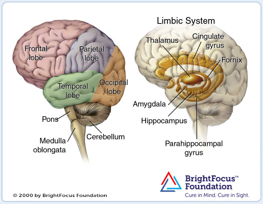
Here is a brief overview of the parts of the human brain and a brain with Alzheimer’s disease.
© 2000 - 2012 American Health Assistance Foundation
The image on the left is a side view of the outside of the brain, showing the major lobes (frontal, parietal, temporal and occipital) and the brain stem structures (pons, medulla oblongata and cerebellum).
The image on the right is a side view showing the location of the limbic system inside the brain. The limbic system consists of a number of structures, including the fornix, hippocampus, cingulate gyrus, amygdala, the parahippocampal gyrus and parts of the thalamus. The hippocampus is one of the first areas affected by Alzheimer’s disease.
Brain With Alzheimer’s Disease
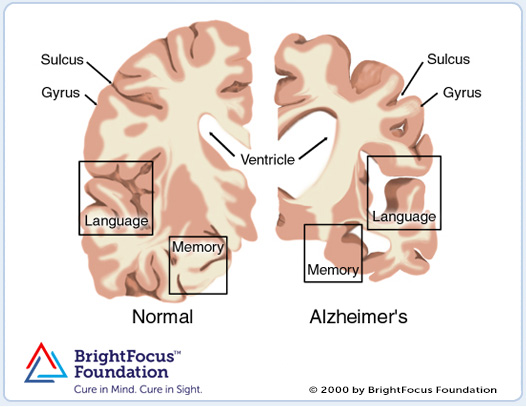
These images represent a cross-section of the brain as seen from the front. The cross-section on the left represents a normal brain and the one on the right represents a brain with Alzheimer’s disease. © 2000 - 2012 American Health Assistance Foundation
In Alzheimer’s disease, there is an overall shrinkage of brain tissue. The grooves or furrows in the brain, called sulci (plural of sulcus), are noticeably widened and there is shrinkage of the gyri (plural of gyrus), the well-developed folds of the brain’s outer layer.
In addition, the ventricles, or chambers within the brain that contain cerebrospinal fluid, are noticeably enlarged. In the early stages of Alzheimer’s disease, short-term memory begins to fade (see box labeled ‘memory’) when the cells in the hippocampus, which is part of the limbic system, degenerate.
The ability to perform routine tasks also declines. As Alzheimer’s disease spreads through the cerebral cortex (the outer layer of the brain), judgment declines, emotional outbursts may occur and language is impaired. As the disease progresses, more nerve cells die, leading to changes in behavior, such as wandering and agitation.
In the final stages of the disease, people may lose the ability to recognize faces and communicate; they normally cannot control bodily functions and require constant care. On average, the disease lasts for 8 to 10 years, but individuals with Alzheimer’s can live for up to 20 years.
Glossary of Terms for an Anatomy of the Brain
Amygdala – limbic structure involved in many brain functions, including emotion, learning and memory. It is part of a system that processes “reflexive” emotions like fear and anxiety.
Cerebellum – governs movement.
Cingulate gyrus – plays a role in processing conscious emotional experience.
Fornix – an arch-like structure that connects the hippocampus to other parts of the limbic system.
Frontal lobe – helps control skilled muscle movements, mood, planning for the future, setting goals and judging priorities.
Hippocampus – plays a significant role in the formation of long-term memories.
Medulla oblongata – contains centers for the control of vital processes such as heart rate, respiration, blood pressure, and swallowing.
Limbic system – a group of interconnected structures that mediate emotions, learning and memory.
Occipital lobe – helps process visual information.
Parahippocampal gyrus – an important connecting pathway of the limbic system.
Parietal lobe – receives and processes information about temperature, taste, touch, and movement coming from the rest of the body. Reading and arithmetic are also processed in this region.
Pons – contains centers for the control of vital processes, including respiration and cardiovascular functions. It also is involved in the coordination of eye movements and balance.
Temporal lobe – processes hearing, memory and language functions.
Thalamus – a major relay station between the senses and the cortex (the outer layer of the brain consisting of the parietal, occipital, frontal and temporal lobes).

American Health Assistance Foundation/ Alzheimer’s Disease Research
Last Review: 01/10/12
http://www.ahaf.org/alzheimers/about/understanding/anatomy-of-the-brain.html
http://www.ahaf.org/alzheimers/about/understanding/brain-with-alzheimers.html
© 2000 - 2012 American Health Assistance Foundation. All rights reserved.